 Open Access



 Open access
 Check for updates


Abstract
As Bangladesh is enriched with fish resources of fresh water, few studies have been conducted to reveal the nutritional value of fish oils. In our present work, we aimed to study the composition of extracted lipids from Colisa fasciatus to explore the medicinal value of this fish which can be used as a precursor for nutraceuticals also. The lipid was extracted from the fish using a solvent extraction method with chloroform and methanol. The qualitative parameters like acid value, peroxide value, saponification value and iodine value were calculated by chemical reaction methods. GCMS analysis was carried out to explore the composition of the extracted lipid. The antioxidant activity of the extracted lipid was carried out by DPPH radical scavenging and Iron reducing study. Thrombolytic activity of the sample was studied by weight difference method. Among 20 constituents hexadecanoic acid (42.808%) was present in the highest concentration. The other remarkable components are Methyl tetradecanoate, pentadecanoic acid, heptadecanoic acid and 9-Octadecanoic acid. Compared to butylated hydroxyl toluene (BHT) as standard, our extracted fish lipid showed 27.9% removal of DPPH. The thrombolytic property showed 7.07 % lysis of blood clots. The fatty acid composition of the extracted lipid supports physiological properties like antiaging and thrombolysis which can make a way to find a new array to use this fish lipid as a new nutritional supplement.
Introduction
Oxidative stress can induce endothelial cell damage, which is a critical event in the initiation of thrombosis. Endothelial cells line the interior surface of blood vessels, and their dysfunction can lead to a pro-thrombotic state. When these cells are damaged by ROS, they can promote the adhesion and aggregation of platelets, leading to clot formation [1]. Additionally, oxidative stress can activate platelets and leukocytes, further contributing to the thrombotic process by releasing pro-inflammatory and pro-coagulant substances [2]. Fatty acids help reduce oxidative stress by neutralizing free radicals, thus preventing cell damage and lowering the risk of chronic diseases. These fatty acids are known to regulate the antioxidant signaling pathways and reduce inflammation, which is a major contributor to chronic non-communicable diseases like cardiovascular diseases (CVDs) and metabolic disorder [3]. Omega-3 PUFAs, including eicosapentaenoic acid (EPA) and docosahexaenoic acid (DHA), have demonstrated strong antioxidant capabilities. Omega-3 PUFAs, including eicosapentaenoic acid (EPA) and docosahexaenoic acid (DHA), have demonstrated strong antioxidant capabilities. They help reduce oxidative stress by neutralizing free radicals, thus preventing cell damage and lowering the risk of chronic diseases. These fatty acids are known to regulate the antioxidant signaling pathways and reduce inflammation, which is a major contributor to chronic non-communicable diseases like cardiovascular diseases (CVDs) and metabolic disorders. They influence platelet aggregation and blood clotting mechanisms, which helps reduce the risk of thrombosis. By altering the production of thromboxanes and leukotrienes, omega-3 fatty acids support platelet homeostasis and prevent excessive clot formation.
This property is particularly beneficial for cardiovascular health, as it can lower the incidence of heart attacks and strokes [4]. These fatty acids are also implicated in improving cognitive function, reducing the severity of inflammatory diseases, and supporting overall immune health. Clinical trials and observational studies suggest that increased intake of EPA and DHA, either through diet or supplementation, can lead to significant health improvements.
Bangladesh has very rich fishery resources both marine and fresh water. Favourable geographical condition, canals, beels, haors, baors, tanks, ponds, ditches etc. have given a unique opportunity for development of expansion of fish industry. About 90% of total demand of the fish in the country comes from fresh water. Because of their high protein content, abundance of vital minerals, and favorable fatty acid profile, fish have long been acknowledged as an important part of human diets. The wide variety of fish species found in diverse environments results in a wide variety of nutritional profiles. For example, fish from freshwater and marine environments have differing fatty acid and bioactive component compositions. As people have started paying more attention to their health, the role of fats in human diet has become more important. Hormone production, cellular membranes, signaling molecules, and energy production are all heavily reliant on lipids [5]. There are three types of fatty acids based on the quantity and existence of double bonds: saturated fatty acids, monounsaturated fatty acids, and polyunsaturated fatty acids (PUFA). Essential fatty acids (EFAs), such as n-3 and n-6 polyunsaturated fatty acids (PUFA), must be obtained from food instead of being synthesized by the human body. The n-6 fatty acids found in current diets are linoleic acid (LA) from foods like soy and corn oil, and arachidonic acid (AA) mostly from meat. However, eicosapentaenoic acid (EPA) and docosahexaenoic acid (DHA), which are mostly found in fish products and are vital for human health, can be converted from alpha-linolenic acid (ALA) in meat [6].
Colisa fasciatus (Family: Anabantidae) is a small subsistence food fish with an indigenous flavor and taste that can be found abundantly in the freshwater habitats in Bangladesh and is renowned by the locals for its medicinal value. They provide a rich resource of proteins, lipids, calcium, vitamin A, and iron in palatable forms in greater abundance than other large freshwater fish [7]. Colisa fasciatus has been revealed as a enriched source of minerals and free fatty acids [7],[8].
In present work, we have aimed to extract the crude lipid from the flesh of Colisa fasciatus and evaluate the quality of the extracted lipid. The extracted lipid has been characterized by GCMS and the lipid has been evaluated as antioxidant and throbolytic agents reported for the first time in this work.
Materials and Methods
Materials
Methanol, Chloroform, Sulfuric acid, Sodium thiosulphate, methanol (GC grade), DPPH, Butylated hydroxyl toluene (BHT), streptokinase were used as reagents and standard. A microprocessor-based pH meter (Model HI 2002 (Edge pH meter, Henna Instruments, UK)) was used for all pH readings. An UV/visible spectrophotometer (T60UV-Visble Spectrophotometer, PG Instruments Limited, UK) was used to measure absorbance. GCMS (Simadzu, Japan) was used for GCMS analysis.
Collection of sample
The Colisa fasciatus, local name: khalisa were purchased from local market in Rajshahi city in Bangladesh. The fishes were washed properly with running water and the flesh was separated from bones. The fish tissue was grounded in pestle mortar and made them paste.
Extraction of total lipid
According to the method of Lee et al. [9] a mixture of chloroform and methanol (2:1 v/v) was used. The pulp was transferred to a volumetric flask (1000 mL capacity) and 600 ml of chloroform-methanol mixture (2:1 v/v) was added and mixed well. For complete extraction it was kept for overnight at room temperature, preferably in the dark. At the end of this period, a further addition of 400 ml chloroform and 400 ml water was mixed. The resulting solution was filtered out then taken in a separating funnel for 30 minutes for phase separation. Three layers are seen, A clear layer of chloroform contained all the lipids, a coloured aqueous layer of methanol with all the water soluble materials and thick pasty interference were seen.
The methanol layer was discarded and the lower layer was carefully collected free of interphase either sucking out with a fine capillary or by filtration through glass wool. The organic layer of the extraction method was taken in a pre weighted round bottom flask for distillation. The mixture is heated to near 60 degree celsius. The chloroform evaporated pass through the distillation column and taken in a volumetric flask for further use.
The sample was covered with a dark paper to protect from light because some lipids got polymerized and decomposed in exposure to light, heat and oxygen. After distillation, the weight of the round bottom flask was taken again. The difference in weight gives the amount of the lipid.
Chemical characterization of extracted lipid
Saponification value determination
The lipid 0.5 gm was taken in a conical flask and 25 mL of alcoholic potassium hydroxide solution was added to it. The flask was then connected to a reflux condenser and heated on a boiling water bath so that the alcoholic solution boiled gently for 30 minute. During this time the flask with its content was shaken occasionally to prevent agitation. A blank experiment (without lipid) was also performed simultaneously in exactly the same manner as described above.
After 30 minutes both the flaks were removed from the water bath and their content, while still hot, were titrated with hydrochloride acid (0.769 N) using phenolphthalein as indicator.
A = The no. of ml of acid required for the blank experiment
B = The no. of ml of acid required for actual experiment.
W = Weight of the lipid taken in gm.
56.1= A constant quantity which represents the gm molecular weight of potassium hydroxide.
Determination of peroxide value
The lipid (1.357 gm.) was taken in a 250 mL glass stopper bottle and 30 mL of acetic acid - chloroform (1:1) was added to it. To the content of the bottle, 0.5 mL of saturated potassium iodide solution was added and the mixture was allowed to stand with occasional shaking. Distilled water (30 mL) was added to the mixture and the content of the bottle was titrated with 0.01 N sodium thiosulfate solution using starch solution as indicator.
A blank experiment (without lipid) was performed exactly in the same manner as described above.
P.V = Peroxide value
S= Volume of mL of sodium thiosulfate solution required for the sample titration
B= Volume of mL of sodium thiosulfate solution required for the blank titration.
N= Normality of sodium thiosulfate solution
Determination of acid value ( as oleic)
A known weight of the lipid (0.532 gm.) was taken in a 25 mL conical flask and mixed with 50 mL of 95% neutralized alcohol. The mixture was heated to boiling and the content of the flask was titrated with aqueous potassium hydroxide solution until a faint pink color persisted for at least 10 second. The content of the flask was shaken continuously and vigorously during the titration. Acid value of the lipid was calculated using the formula given below [11]:
Determination of ester value [12]
For the oil or lipid to be analyzed, the acid value and the saponification value were determined which had been described before. The ester value was determined using the formula,
Determination of iodine value (HENUS METHOD)
The lipid (0.537 gm) was dissolved in 10 mL of chloroform in a dry glass stoppered bottle (500 mL). To the content of the bottle 25 mL of Henus solution was added and the mixture was allowed to stand in the dark for exactly 30 minutes with occasional shaking. Potassium iodide solution (10 mL) was mixed to it and the mixture was shaken well. Freshly boiled and cooled distilled water (100 mL) was added to the mixture and the content of the bottle was titrated with standardized sodium thiosulfate (0.097 N), using starch solution as indicator [13].
A blank experiment (without the lipid) was performed exactly in the same manner as described above.
GC-MS Anaysis of extracted lipid from Colisa fasciatus
Gas chromatography analysis was performed using a GC-2010 Plus Shimadzu fitted with GCMS-QP2020 Shimadzu. Column length 30m (L) X 0.25mm (ID), X 0.25um (DF). The total running time was 32.3 minutes. Helium was the carrier gas while the injection volume was 1 μL. The injection port was maintained at 250ºC, and the split ratio was 75:1. Oven temperature programming was done from 140 to 250ºC at 10ºC/min, and it was kept at 250ºC for 10 min. The spectra of the compounds were matched with NIST and Wiley library. The structures were defined by the % similarity values and confirmed by the study of classical fragmentation pattern, base peak and molecular ion peaks of the compounds.
Antioxidant activity of extracted lipid
DPPH Radical Scavenging Assay
Stock solution of extract and BHT was prepared (0.4 mg/mL). Five screw cap tubes were labeled as 10, 20, 40, 80, 160, µg/mL. 125, 250, 500, 1000, 2000 µL stock solution of extract and BHT was taken on that consecutive labeled test tube and then add required amount of methanol to make the final volume of 2 ml, using micropipette. Three milliliters of this DPPH solution was mixed with 2 mL of extract solution and standard solution separately. These solution mixtures were kept in dark for 30 min. The degree of DPPH purple decolorization to DPPH yellow indicated the scavenging efficiency of the extract. The absorbance of DPPH solution (Control solution ‘A’) was measured at 517 nm using UV Visible Spectrophotometer. The absorbance of the mixture was also determined at 517 nm using UV-Visible Spectrophotometer. BHT was served as standard. Lower absorbance of the reaction mixture indicated higher free radical-scavenging activity
A is the absorbance of control (DPPH solution without the sample),
B is the absorbance of DPPH solution in the presence of the sample (extract/ BHT).
Iron reducing activity
1 mL sample and standard solution at various concentrations (10, 20, 40, 80, 160 μg/mL), 2.5 mL of potassium buffer (0.2M) and 2.5 mL of 1% potassium ferricyanide, [K3 Fe (CN)6] solution were added into the test tubes. The reaction mixtures were incubated for 20 min at 50°C to complete the reaction. Then 2.5 mL of 10% trichloro acetic acid (TCA) solution was added into the test tubes. The total mixture was centrifuged at 3000 rpm for 10 min, after which 2.5 mL supernatant was withdrawn from the test tubes and mixed with 2.5 mL of distilled water and 0.5 mL of 0.1% ferric chloride (FeCl3) solution. The absorbance of the solution was measured at 700 nm using a spectrophotometer against blank. A typical blank solution contained the same solution mixture without plant extracts/standard and was incubated under the identical conditions. The absorbance of the blank solution was measured at 700 nm.Increasing absorbance of the sample indicates the incresing iron reducing activity.
Thrombolytic activity
4 mL venous blood drawn from chicken was distributed in three different pre weighed sterile micro centrifuge tubes (0.5mL/tube) and incubated at 37°C for 45 minutes. After clot formation, serum was completely removed without disturbing the clot and each tube having clot was again weighed to determine the clot weight (clot weight = weight of clot containing tube – weight of tube alone). To one micro centrifuge tube containing pre-weighed clot, 100 µL of aqueous extract of Colisa fasciata added. As a negative non thrombolytic control, 100 µL of distilled water were separately added to the control tubes numbered. All the tubes were then incubated at 370C for 90 minutes and observed for clot lysis. After incubation, fluid released was removed and tubes were again weighed to observe the difference in weight after clot disruption. Difference obtained in weight taken before and after clot lysis was expressed as percentage of clot lysis.
Result and Discussion
Qualitative parameter analysis of extracted lipid
The parameters e.g saponification value, peroxide value, acid value, ester value and iodine value refers to characterization of lipid. These parameters estimated in our present work have been represented in table 1.
| sample | Saponification value | Peroxide value | Acid value | Ester value | Iodine value |
|---|---|---|---|---|---|
| Extracted Lipid | 295.544 | 1.47 | 1.62 | 188.12 | 51.35 |
GCMS Analysis of extracted lipid
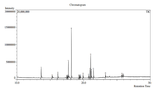
The sample were analysed, GC-MS chromatogram of fatty acid methyl ester mixture is given in (Fig. 1) and the fatty acid composition of the methyl ester mixture derived from the lipid of Colisa fasciata is given in (table 2). It is evident from the table that the lipid of Colisa fasciata contained highest amount of Hexadecanoic acid (42.80%), Methyl tetradecanoate (12.63%), 9-Octadecenoic acid (z) (8.52%), Pentadecanoic acid (5.26%), 9-Hexadecanoic acid (6.17%), 4,7,10,13,16,19-Docosahexaenoic acid (0.74%), Arachidonic acid (0.996%) were prominently present in our sample.
Antioxidant activity of the extracted lipid
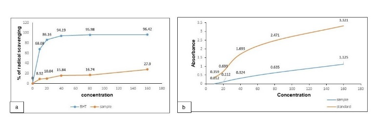
DPPH Scavenging Study: The DPPH radical scavenging activity of the extracted lipid was presented in the Fig. 2. The graph showed that the lipid was able to reduce the DPPH free radicals n the gradient concentration manner. The lipid showed 27.9% removal of free radicals at 160 g/mL. The iron reducing activity was carried out compared to BHT as standard. The method was continued from 0 to 160 g/mL. The absorbance was increased gradually with the increase of concentration that indicates the potential antioxidant activity of our sample.
Thrombolytic activity
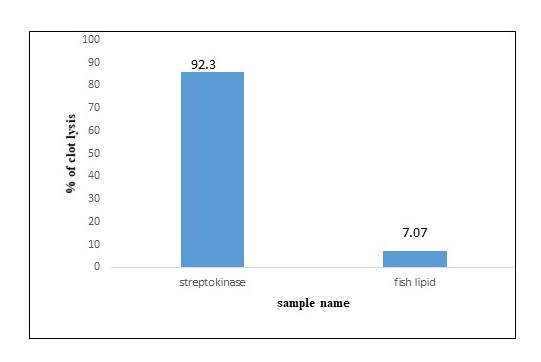
The sample was presented as clot lytic agent which showed 7.07% of thrombolysis compared to streptokinase as standard. The found data was accumulated in the figure 3.
As fatty acid composition of lipid from marine fish has been explored in several cases, there is lack of investigation on lipid extracted from fresh water fish. In our present work we have selected one of fresh water fishes abundant in Bangladesh so that the quality and the composition of the extracted lipid of the respective fish can be evaluated to explore the nutritional and pharmacological importance of the fish. Saponification value, acid value, ester value, peroxide value and iodine value have been estimated to study the quality of the extracted lipid. The saponification value was found 295.544 according to our study. Saponification value (SV) is an index of the average molecular mass of fatty acid in the oil sample. Peroxide value refers to the presence of peroxide in lipid that causes rancidity in the oil. When polyunsaturated fatty acid is oxidized, peroxide is formed as a primary free radicals [14]. In our study the peroxide value refers within acceptable limit as it should be less than 5 [15]. In -this study, the fatty acid analysis of extracted lipid revealed the presence of several derivatives of hexadecanoic acid which belongs to be one of the omega 3 members. The omega 6 and omega 3 ratio has been suggested by several researches as a helpful gauge of the nutritional content of fish oil, with a lower ratio being more beneficial in reducing heart illnesses linked to plasma lipid levels [4]. The Food and Agriculture Organisation (2014) states that the ω-6/ω-3 ratios in the human diet shouldn't be more than 5.0.
According to the GCMS analysis, we have found a derivative of Docosahexaenoic acid as 4,7,10,13,16,19-Docosahexanoic acid. Docosahexaenoic acid is a polyunsaturated fatty acid found in fish oil, exerts cytotoxic effects on cancer cells [16]. The octadecanoic acid derivatives and arachidonic acid have been found in our sample which exert free radical removing activity. The antioxidant activity presented in our experiment justify that the free fatty acids present in the extracted fish lipid may contribute to the property. According to previous report, omega-3s are capable of reducing free radical levels although it is not considered as a super antioxidant [17]. It is important to note that both in vitro and in vivo studies have indicated that PUFA consumption reduces the excretion of lipid peroxidation products and that ω3 FAs supplements do not increase the production of reactive oxygen species. Therefore, in vascular endothelial cells, ω3 FAs may function indirectly as antioxidants rather than pro-oxidants, reducing inflammation and subsequently the risk of atherosclerosis and cardiovascular illnesses. In our study we found thrombolytic activity of the fish lipid although it cannot be regarded as prominent. The antioxidant compounds may contribute to the blood clotting as the mechanism on the haemostatic system, in particular on blood platelets and endothelium.by omega 3 has been reported earilier [18] . Previous researches have revealed that lipids extracted from fish exhibited PAF-inhibiting activities. Given that PAF is an initial trigger in atherosclerosis [19].
Conclusion
We conducted the study as preliminary attempt to explore the fatty acid composition of the lipid from one of the available fresh water fish named Colisa fasciata. The qualitative parameters of the respective fish lipid revealed the lipid as promising polyunsaturated fatty acid. The presence of omega3 precursor and many other polyunsaturated fatty acids as well as the biological responses of the extracted lipid has revealed it as one of the promising natural resources for functional supplements. Further studies are recommended to develop supplement of fatty acid from this resource.
Declarations
- Author contributions
Md Tamzid Hossain Molla: Conceptualization, data analysis, supervision, review and editing. Shajid Hasan: Formal analysis, methodology, data curation, writing draft; Mahci Al Bashera: Resources, software, writing draft, review and editing; Bipasha Kabir: Health threat mitigation, logistic support and Ahmed Tareq Shams Chowdhury: Experimental analysis.
- Funding
The whole research has been conducted with no financial grants or funding and logistic support from any institution and organisation.
- Data availability
Not applicable
- Competing interests
Not applicable
- Acknowledgments
We would like to acknowledge the Department of Applied Chemistry and Chemical Engineering, University of Rajshahi for providing laboratory facilities. We are also grateful to BCSIR, Rajshahi Laboratory to provide the facilities to conduct the GCMS analysis of our present work.
References
- 1.Chaudhary, P., Janmeda, P., Docea, A. O., Yeskaliyeva, B., Abdull, A. F. R., Modu, B., Calina, D. and Sharifi-Rad, J. (2023). Oxidative stress, free radicals and antioxidants: potential crosstalk in the pathophysiology of human diseases. Frontiers in Chemistry, 11: 1–24. doi: https://doi.org/10.3389/fchem.2023.1158198.
- 2.Lichota, A., Szewczyk, E. M., Gwozdzinski, K. (2020). Factors Affecting the Formation and Treatment of Thrombosis by Natural and Synthetic Compounds. International Journal of Molecular Sciences, 21(21). doi: https://doi.org/10.3390/ijms21217975.
- 3.Lorente-Cebrián, S., Costa, A. G. V., Navas-Carretero, S., Zabala, M., Laiglesia, L. M., Martínez, J. A., Moreno-Aliaga, M. J. (2015). An update on the role of omega-3 fatty acids on inflammatory and degenerative diseases. Journal of Physiology and Biochemistry, 71(2): 341–9. doi: https://doi.org/10.1007/s13105-015-0395-y.
- 4.Djuricic, I., and Calder, P. C. (2021). Beneficial Outcomes of Omega-6 and Omega-3 Polyunsaturated Fatty Acids on Human Health: An Update for 2021. Nutrients, 13(7). doi: https://doi.org/10.3390/nu13072421.
- 5.Dyall, S. C. (2015). Long-chain omega-3 fatty acids and the brain: A review of the independent and shared effects of EPA, DPA and DHA. Frontiers in Aging Neuroscience, 7(APR): 1–15. doi: https://doi.org/10.3389/fnagi.2015.00052.
- 6.Taşbozan, O., Gökçe, M. A. (2017). Fatty Acids in Fish. In: Catala A, editor. Fatty Acids. Rijeka: IntechOpen;. Available from: https://doi.org/10.5772/68048
- 7.Islam, R., Hossain, M. B., Islam, M. N., Islam, M. M., and Islam, M. T. (2020). Nutrient composition of small indigenous fish species (SIS) from homestead ponds of Noakhali coast, Bangladesh. Egyptian Journal of Aquatic Biology and Fisheries, 24: 943–954. doi: https://doi.org/10.21608/EJABF.2020.133693.
- 8.Sumi, K., Sharker, R., Rubel, M., and Islam, M. (2023). Nutritional composition of available freshwater fish species from homestead ponds of Patuakhali, Bangladesh. Food Chemistry Advances, 3:100454. doi: https://doi.org/10.1016/j.focha.2023.100454.
- 9.Lee, C. M., Trevino, B., and Chaiyawat, M. (1995) Simple and Rapid Solvent Extraction Method for Determining Total Lipids in Fish Tissue. Journal of AOAC International, 79: 487–92. doi: https://doi.org/10.1093/jaoac/79.2.487.
- 10.Stansby M., (2002) Determination of Peroxide Values for Rancidity in Fish Oils. Industrial & Engineering Chemistry Analytical Edition, 13(9): 627-631. doi: https://doi.org/10.1021/i560097a017.
- 11.Islam, M., Naznin, S., Islam, M. R., and Rahman, M. (2019). Extraction and Characterization of Lipid from Pangus Fish (P. Pangasius), Available in Bangladesh by Solvent Extraction Method. American Journal of Zoology, 1(2), 28-34. doi: https://doi.org/10.11648/j.ajz.20180102.11.
- 12.Alajtal, A. I., Sherami, F. E., and Elbagermi, M. A. (2018). Acid, Peroxide, Ester and Saponification Values for Some Vegetable Oils Before and After Frying. American Associstion for Science and Technology, 4(2): 43–47.
- 13.American Society for Testing Materials. Standard Test Method for determination of the Saponification Value of Fats and Oils, ASTM (D 5558 – 95). 2001;95(Reapproved):1–2.
- 14.GOED (2015), Nutrition C of R. Oxidation in Omega-3 Oils : An Overview.;8. Available from: https://drive.google.com/file/d/0B6uJWj5y9FY9Q3hUeUdYcExxTjA/view?pref=2&pli=1
- 15.Jairoun, A. A., Shahwan, M., and Zyoud, S. H. (2020). Fish oil supplements, oxidative status, and compliance behaviour: Regulatory challenges and opportunities. PLoS One, 15(12): 1–12. doi: https://doi.org/10.1371/journal.pone.0244688.
- 16.Bégin, M. E., Ells, G., and Horrobin, D. F. (1988). Polyunsaturated fatty acid-induced cytotoxicity against tumor cells and its relationship to lipid peroxidation. Journal of the National Cancer Institute, 80(3): 188–194. doi: https://doi.org/10.1093/jnci/80.3.188.
- 17.Akmal, N. L. H. B. I., and Roy, A. (2017). Free Radical Scavenging Activity of Fish Oil-an in-Vitro Study. International Journal of Pharmaceutical Sciences and Research, 8(9): 3872–3875. doi: https://doi.org/10.13040/IJPSR.0975-8232.8(9).3872-75.
- 18.Golanski, J., Szymanska, P., and Rozalski, M. (2021) Effects of omega-3 polyunsaturated fatty acids and their metabolites on haemostasis—current perspectives in cardiovascular disease. International Journal of Molecular Sciences, 22(5): 1–14. doi: https://doi.org/10.3390/ijms22052394.
- 19.Wang, C., Xia, W., Xu, Y., Jiang, Q., Yin, S., Yang, Y, and Yu, P. (2013). Anti-platelet-activating factor, antibacterial, and antiradical activities of lipids extract from silver carp brain. Lipids in Health and Disease, 12: 1-6. doi: https://doi.org/10.1186/1476-511X-12-94
Cite this article
Journal
Molla MTH, Hasan S, Bashera MA, Kabir B, Chowdhury ATS. Fatty Acid Composition, Antioxidant Activity and Thrombolytic Activity Analysis of Extracted Lipid from Colisa fasciatus. JoSEP. 2026;1(2):90-95. doi: 10.62275/josep.24.1000013.
Rights and permissions
© 2026
Cited by (1)
- Native Collagen and Total Lipid Extract Obtained from Caranx hyppos By-Products: Characterization for Potential Use in the Biomedical and Nutraceutical Fields
Sheyza Menéndez-Tasé, Evelin Gaeta-Leal, Darío Iker Téllez‐Medina · Marine Drugs · 2025
About this article
- Received
- 2 August 2024
- Revised
- 17 August 2024
- Accepted
- 18 August 2024
- Published
- 21 October 2026
- Journal
- Journal of Science and Engineering Papers
- Volume / Issue
- 1 / 2
- Pages
- 90-95
- DOI
- 10.62275/josep.24.1000013