 Open Access



 Open access
 Check for updates


Abstract
Rice husk is an abundant natural resource that contains about 33% cellulose. This study focused on the extraction and develop of carboxymethyl cellulose (CMC) from rice husks and its utilization in developing CMC-polyvinylpyrrolidone (PVP) blended hydrogel (HGEL) membranes with varying CMC:PVP ratios (80:20, 60:40, and 50:50 w/w). The controlled release of pharmaceuticals and targeted drug administration depend on pH-responsive drug delivery systems. An innovative pH-sensitive, biodegradable, and antibacterial hydrogel was synthesized through blending and solution casting method, using glutaraldehyde as a crosslinking agent. Structural and physicochemical characterization confirmed successful hydrogel formation: FTIR validated crosslinked network development, TGA demonstrated enhanced thermal stability, and SEM revealed uniform and interconnected morphologies. Swelling studies showed that swelling capacity could be precisely tuned by altering the CMC/PVP composition, with higher pH conditions markedly increasing swelling, confirming the pH-responsive behavior of the hydrogels. The drug (salicylic acid) release profiles from gel membrane were conducted into two separate release media (pH 1.4, and pH 7.4), with samples evaluated spectrophotometric analysis at 294 nm wavelength using a UV-Vis spectrophotometer, showed a consistent and controlled release profile. Biodegradability tests in soil revealed that the hydrogel sample degraded gradually up to 72.65% within 15 days. Moreover, upon testing on bacteria Staphylococcus aureus (G+) & Escherichia coli (G-) bacterium, the sample (50:50) gave 11 mm of inhibition zone diameter which confirmed that the synthesized hydrogel can be used for wound dressing and other biomedical applications. Altogether hydrogels underlined as a prospective category of eco-friendly and tunable biomaterials for future healthcare innovations.
Introduction
Hydrogels are unique three-dimensional networks of polymers, which can absorb and hold considerable water or biological fluids without dissolution because polymeric chains are physically or chemically crosslinked [1], [2]. In the hydrated state, hydrogels are soft, elastic and rubber-like material-similar to natural living tissue-and thus attract considerable interest in pharmaceutical and biomedical applications. Drug delivery systems are of such interest, as it is possible to encapsulate therapeutic agents inside the hydrogels and deliver them in a controlled and predictable manner [3].
Traditional drug delivery methods often have some deficiencies including premature release, inefficiency of localization, toxicity to the whole system and low bioavailability. To overcome these limitations, the stimuli-responsive hydrogels have become attractive drug carriers for responding to physiological stimulations such as pH, temperature, ionic strength and enzymatic activity [4]. Among these stimulation, pH-sensitive hydrogels are especially important as natural changes in pH occur throughout the gastrointestinal tract in addition to inflamed tissues, infected wounds and the tumor microenvironments. Thus, pH-responsive hydrogel that can swell and shrink in response to pH change makes it possible to achieve site-specific and sustained drug release, and minimize the side effects also maximizing therapeutic efficacy [5], [6].
Because of the abundant resource, renewability, biodegradability and good biocompatibility, cellulose-based polymers have attracted extensive attention for fabricating the drug delivery hydrogel [7]. Carboxymethyl cellulose (CMC), a water-soluble anionic derivative of cellulose, is a cereal gum that has been extensively used in pharmaceutical and biomedical applications because of its non-toxicity, hydrophilicity, film-forming ability, and pH-sensitive properties [8]. As carboxyl (-COOH) groups are present in CMC, it is possible to ionize under various pH conditions, which leads to pH-dependent swelling and drug release properties of CMC hydrogels. Therefore, CMC-based hydrogels have been extensively investigated as carriers for oral, transdermal and wound-healing drug delivery systems [9], [10].
Nevertheless, CMC based hydrogels have several drawbacks such as poor mechanical properties, excessive swelling and poor structural stability, which makes them unsuitable for practical uses [11]. To overcome these challenges, combining CMC with biocompatible synthetic polymers for which crosslinking agents can be added is a successful approach. Polyvinylpyrrolidone (PVP) is non-ionic water-soluble biocompatible synthetic polymer extensively used in pharmaceutical formulation as binding agent, stabilizer, plasma expander and drug carrier [12], [13]. In case of PVP, lactam groups in PVP molecules can be able to hydrogen bond with polymer- and drug-matrix, which may enhance the mechanical properties and control drug release behavior [14]. The combination of CMC and PVP in hydrogel matrices provides several advantages such as augmented mechanical strength, improved regulation over swelling behavior and controlled drug release patterns [15]. In addition, such host-guest interaction between natural and synthetic polymers gives the advantage of combining biocompatibility and biodegradability from CMC with the flexibility and structural stability of PVP leading to hydrogels that have better performance than their single component counter parts [16]. These polymers mixed hydrogels are especially attractive materials for the construction of pH-responsive drug delivery systems.
Crosslinking is an important property and it defines the physicochemical properties of hydrogels such as swelling, porosity, mechanical strength and degradation [17]. Glutaraldehyde is one of the most popular chemical cross-linking agents, leading to the formation of stable three-dimensional networks [18], [19]. High concentrations of glutaraldehyde may be toxic to the cells; however, controlled treatments have been shown to result in mechanically robust hydrogels that are useful for biomedical applications. The thermally stable, structurally intact hydrogels did not collapse and reached a swelling equilibrium in time during control-release experiments, necessary for ultimate release formulations or storage [20].
Salicylic acid is a widely used pharmaceutical compound for various diseases, for example: anti-inflammatory, keratolytic, and antimicrobial treatment. However, topical salicylic acid delivery directly is frequently characterized by burst release, skin irritation, and lack of control over the therapeutic process [4], [21]. As a result of incorporating the SA into a hydrogel matrix, we found it to be used effectively for controlling release rate, preserving its chemical stability and improving therapeutic efficacy. pH-sensitive hydrogels are well suited for delivering salicylic acid, and the responsive degree of swelling of such a gel and drug diffusivity both depend on pH change [22], [23].
Apart from controlled drug release, biodegradability and antimicrobial action are highly desired properties of hydrogel systems for biomedical usage in vivo including wound healing and tissue regeneration [24], [25]. As reported, cellulose-based hydrogel degrades slowly in natural and biological environment can reduce long term toxicity and environmental pollution. Also, the incorporation of antimicrobials or medications in hydrogels may exhibit localized antimicrobial effects for infection control and healing enhancement [26].
In this study, blending with CMC using modified mixing method is the selected method in fabrication pH responsive membranes and PVP was selected because of its anti-tumorigenic properties in drug delivery and glutaraldehyde used as a cross linking agent. The effects of several CMC:PVP proportions on the structural, thermal, morphological, swelling, degradation and drug release behavior of the synthesized hydrogels were also studied. Salicylic acid was chosen as a model drug in order to study the pH-responsive release properties of the hydrogel membranes under simulated physiological conditions. The findings indicate that the fabricated CMC-PVP hydrogels have controlled and sustained drug release, significant pH-responsiveness, biodegradation ability and antimicrobial properties making them significant alternative for efficient drug delivery systems in biomedical and wound healing applications.
Materials and methods
Materials
Rice husk was collected from a local rice mill in Bangladesh and used as the raw biomass material. Sodium hydroxide pellets (NaOH, 98.7% of purity), glacial acetic acid (CH3COOH, 99% of purity), absolute ethanol (C2H5OH, 99.5% of purity), and hydrochloric acid (HCl, 37% of purity) were purchased from Merck, Darmstadt, Germany. Sodium chlorite (NaClO2) and sulfuric acid (H2SO4) were used as received. Monochloroacetic acid (CH2ClCOOH, 99% of purity) was obtained from Qualikems Fine Chem Pvt. Ltd., India. Polyvinylpyrrolidone (C6H9NO)x was supplied by Sisco Research Laboratories Pvt. Ltd., Mumbai, India, while glutaraldehyde (C5H8O2, 25% of purity) was procured from Finar Ltd., Gujarat, India. Merck in Darmstadt, Germany, provided standard buffer solutions with pH values of 4.0, 7.2, and 9.4. All compounds were analytical grade and utilized without additional purification.
Extraction of cellulose by hydrothermal process
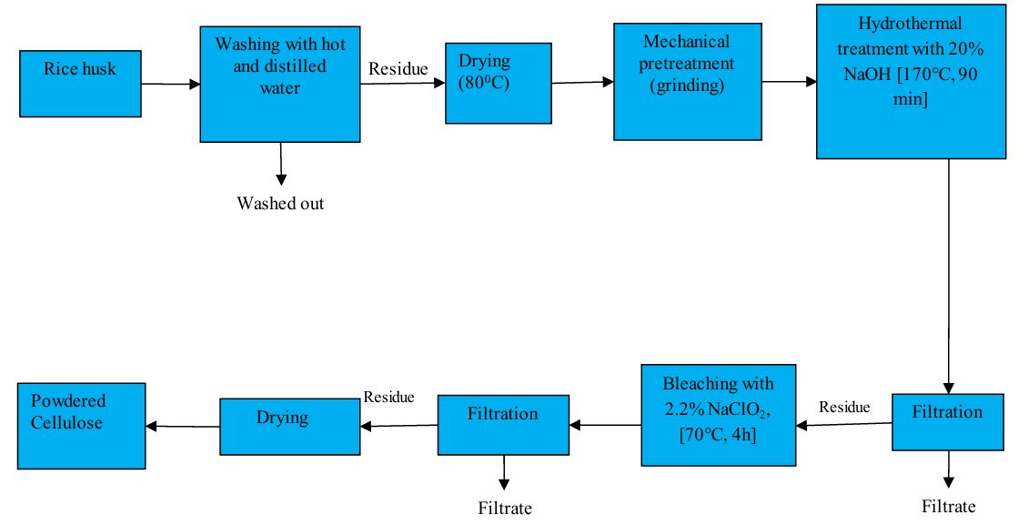
Rice Husk (sample) was collected and washed with hot water and then distilled water, respectively. Then the washed sample was dried into hot air oven at 800C until it become dry and grinded finely thereafter. It was mixed with 20% NaOH in a 250 mL beaker and transferred to a Teflon-lined hydrothermal reactor. The reactor was heated at 170°C for 90 minutes, after which the sample was filtered, washed to neutrality, and dried at 80°C for 5 hours to obtain cellulose residue. The dry cellulose was bleached using a 2.2% sodium chlorite solution at a weight ratio of 1:0.5 powder to sodium chlorite. Acetic acid was added to the solution to reduce its pH to 3.7, followed by a 4-hour reaction at 70°C. The pure cellulose was recovered by filtration, washed repeatedly until it was free of acid, and lastly dried to achieve a constant weight [27]. The total process was repeated twice to four times for better removal of lignin. A schematic diagram of cellulose extraction process from rice husk has been shown in scheme-1.
Synthesis of carboxymethyl cellulose (CMC)
NaOH was added 50 minutes after cellulose was added to a 30% ethanol aqueous solution while being stirred magnetically. At 300ºC, the alkalization (steeping) process was carried out. A 120% (w/v) aqueous monochloroacetic acid solution was added dropwise at 650ºC and agitated for three hours following the alkalization reaction. Carboxymethylation is the term for this process. After neutralizing the solution with HCl, it was filtered. The residue dried until its weight remained constant [28].
Preparation of hydrogel
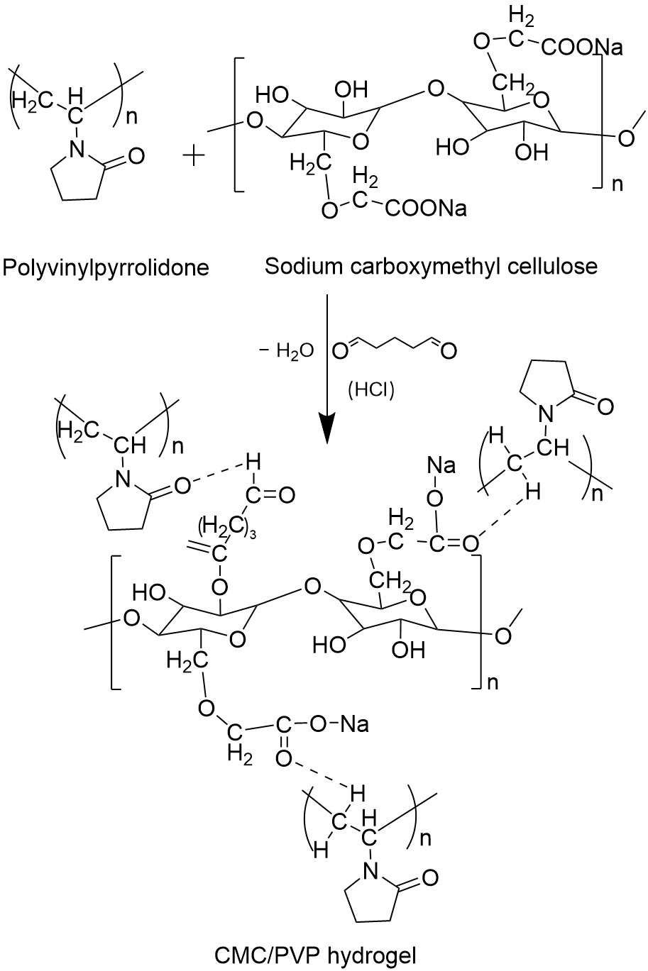
A 20 mL solution of 5% (w/v) CMC in water was prepared in a beaker. To this CMC solution, 5 mL of PVP solution (containing 0.25 and 1 g of PVP to achieve CMC: PVP ratios of 80:20 and 50:50) was added drop wise with continuous stirring to ensure a homogeneous mixture. Glycerin was then added to the CMC-PVP blend while stirring to prevent brittleness in the hydrogel. A glutaraldehyde reagent (HCl + GA) was introduced as a crosslinking agent, and the resulting dispersion was stirred at room temperature for two hours to allow proper reaction between CMC and PVP. The thick dispersions obtained were then converted into hydrogel membranes using conventional methods. The hydrogel membranes were left to dry at room temperature for 72 hours. Once dried, the membranes were peeled off and thoroughly washed with distilled water to remove any residual HCl and glutaraldehyde (GA). The resulting hydrogel membranes were labeled as HGEL-1 and HGEL-2. The resulting hydrogel possible reaction mechanism is given in scheme 2.
Characterization
The Fourier Transform Infrared (FTIR) spectrophotometer-3000 Hyperion Microscope Vertex 80 was used to examine the different functional groups of Na-CMC. FTIR spectra with a resolution of 1 cm−1 were acquired from KBr pressed pellets in the 4000-400 cm−1 range. The morphology (granule surface and shape) of the produced hydrogel was examined using scanning electron microscopy (SEM). Scanning electron microscopy (SEM) was used to examine the morphology of produced cellulose (Model: ZEISS EVO 18).
Determination of water absorption of hydrogel with soaking time
Hydrogel samples after extraction were dried to a constant weight and immersed in distilled water at room temperature. At first, weight of swelled gel was taken 1, 3, 7, 12 and 15 hours and it was continued after 24, 48 and 72 hours. After completion of absorbing period, the hydrogels were taken out and adhering water was removed using tissue paper. It was then weighed. The water absorption (Wa) can be calculated as g H2O/g dry copolymer using the following equation:
Where, m2 is the weight of sample with absorbed water and m1 is the weight of dried sample.
Swelling study at different pH level
The dried hydrogel membranes (HGEL-1 and HGEL-2) were immersed directly in buffer solutions of pH 1.2, 5.4, 7.4 and 9.4 at room temperature for 72 h. The equilibrium percentage of swelling (% swelling) of the product was calculated as
Where WS is the weight of the product after hydration for 72 h and WD is the initial weight of dried hydrogel membrane [29].
Determination of gel fraction in hydrogel
The hydrogel sample was dried to a constant weight. The gel content in the dried sample was estimated by measuring its insoluble part in dried sample after immersion in deionized water for 48h at room temperature [30].
The gel fraction was calculated as follows,
Where, Wi is the initial weight of dry gel and Wd is the weight of dried insoluble part of sample after extraction with water.
Determination of biodegradable properties of hydrogel
Studies on the hydrogel samples' degradation were carried out in sandy and clay soils. Samples were taken out on a regular basis, cleaned with distilled water, and dried until their weight remained consistent. The following formula can be used to determine the hydrogel's biodegradability:
Drug loading of hydrogel
Salicylic acid (SA) was used as a model drug to investigate drug loading in the crosslinked CMC/PVP hydrogel membrane. Diffusion was used to add SA to the hydrogel membrane. The SA solution in acetone (1 g SA in 10 ml acetone) was used to submerge the CMC/PVP hydrogel membrane for five hours. To remove the medication that had adhered to the surface of the membrane, distilled water was used to wash the drug-loaded hydrogel membrane. Drug concentrations were evaluated both before and after the hydrogel membrane was submerged in the staining solution [31].
Drug releasing efficiency of hydrogel
A magnetic stirrer was used to mechanically agitate the drug-loaded CMC/PVP hydrogel membrane (HGEL-2) in a beaker filled with a 100 ml pH 7.4 solution. Five milliliters of releasing medium were collected in the glass vial and replaced with five milliliters of stock solution at equal intervals of twenty minutes. Using the same technique, the drug release investigation was also conducted at SGF (stimulated gastric fluid pH 1.2) and distilled water. Samples were then subjected to spectrophotometric analysis using a UV-Vis spectrophotometer at a wavelength of 294 nm [31].
Assays to determine antimicrobial activity of hydrogel
Antimicrobial activity of the compounds was measured by observing the growth response of Staphylococcus aureus (a gram positive model) and Escherichia coli (a gram negative model). The susceptibility of such growth rate of microorganism was measured in vitro by disc diffusion method [32]. The steps were conducted in a laminar air flow cabinet under aseptic conditions. Freshly developed bacterial cultures were used to create nutrient agar plates, which were then left to solidify. Kanamycin (30 µg/disc) was employed as the reference antibiotic, and sterile (BBL, U.S.A.) filter paper discs (5 mm diameter) impregnated with the test samples were placed on the inoculated agar plates. The plates were incubated for 12–18 hours at 37 °C after being held at 4 °C for diffusion. The diameter of the zones of inhibition (mm) was used to calculate the antimicrobial activity.
Results and discussion
FT-IR Analysis
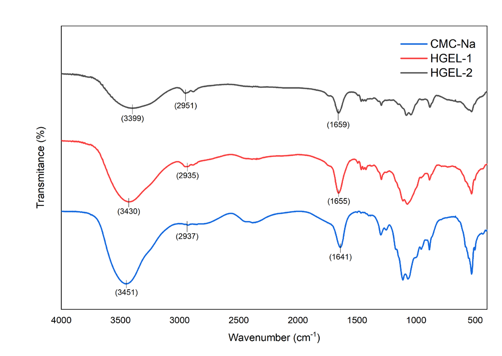
Figure 1 shows the FT-IR peaks for CMC-Na, HGEL-1, and HGEL-2. The FT-IR of CMC-Na reveals a peak at 3451 cm−1, which illustrates the stretching of the –OH group in carboxylic acid. The signal at 2937 cm−1, on the other hand, demonstrated C-H stretching of alkane. The peak for C=O stretching vibrations caused by amide (I) was at 1641 cm−1. The peaks at 1423 and 1329 cm−1 could be due to the bending modes of CH2 and CH3 in alkanes, respectively. The peak at 1142 cm−1 was thought to be caused by C-N stretching vibrations because it had aromatic amine groups. The signal at 1027 cm−1 demonstrated that carboxylic acids were stretching C-O. The signal at 535 cm−1 showed that alkynes C-H bending was present. The FTIR spectra of produced CMC/PVP blended hydrogel membranes (HGEL-1, HGEL-2) exhibit similar peaks between 1650–1660 cm−1, corresponding to amide (I). The creation of hydrogen bonds decreased the strength of the carbonyl bond in PVP in all of the hydrogel membranes and moved the carbonyl stretching to a lower frequency. The FTIR spectra of CMC/PVP hydrogel membranes show that when PVP is mixed with CMC, the –OH stretching vibration peak of CMC at 3451 cm−1 moves to lower frequencies of 3430 and 3399 cm−1. The presence of hydrogen bonding in the hydrogel membranes can explain why the –OH groups' frequency has gone down. The peaks between 2,930 and 2,955 cm−1showed that there was a peak for the –CH stretching vibration. The peaks between 1740 and 1760 cm−1 are caused by the carboxymethyl groups (–CH2OCH2COOH) in the hydrogel membranes. The peaks at 1030 and 1042 cm−1 reveal that the hydrogel membranes have a stretching vibration peak for –CN. The peaks suggest that the hydrogel was made by successfully combining CMC and PVP.
Swelling kinetics in different aspect
The effect of amount of crosslinker on swelling of the hydrogel was studied varying amount of crosslinker (5-14%) in Figure 2. The maximum swelling was obtained at 10% crosslinker with respect to CMC. Above or below this amount of crosslinker, the swelling percent of hydrogel was decreased. Below this amount proper reaction cannot be formed and excess crosslinker can cause strong bond formation in the hydrogel which cause less swelling of hydrogel. From this, hydrogels are synthesized with 10% crosslinker.
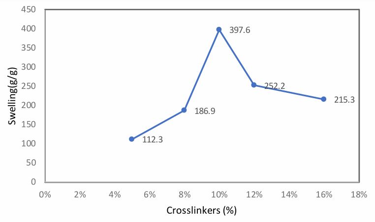
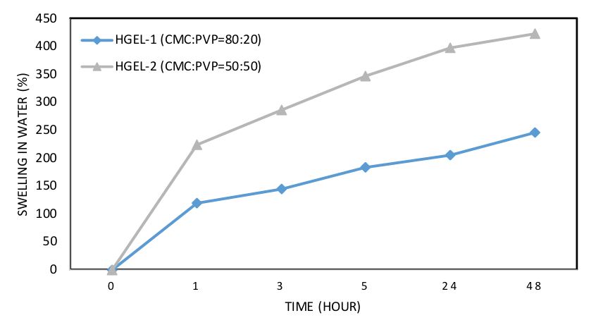
Figure 3 shows how hydrogels in water swell over time. All hydrogel reacted with water in different ways. Over time, the hydrogels showed an increase in swelling. Because more PVP may result in more hydrogen bonding between CMC and PVP, an increase in PVP ratio in membranes raises the swelling ratio of hydrogel. A 50:50 ratio of CMC to PVP can result in the greatest swelling. Because hydrogel has certain hydrophilic groups, it absorbs water. Water was absorbed by it until those groups were saturated. By forming hydrogen bonds with the molecules, it also took up water.
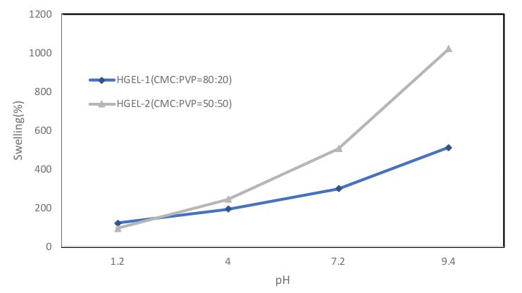
The crosslinked CMC/PVP hydrogel membranes (HGEL-1, HGEL-2) were immersed in buffer solutions with pH values of 1.2, 4, 7.2, and 9.4 to observe their swelling behavior. Figure 4 illustrates the swelling patterns of these hydrogel membranes. The results show that swelling increased with pH for all CMC/PVP hydrogel compositions (HGEL-1, HGEL-2). The maximum swelling degree was observed at pH 9.4 (513%, 1022%), in contrast to the swelling at pH 1.2. Since the apparent pKa value of CMC is 4.756, at pH values lower than this, the carboxymethyl groups (–CH2COOH) of CMC collapse, leading to a lower swelling degree. Additionally, hydrogen bonding occurs between the hydroxyl group (–OH) of CMC and the carbonyl group (–C=O) of PVP, which further reduces swelling at these lower pH values. However, at higher pH values (above the pKa value), the hydrogel's swelling increases due to the dissociation of the hydrogen bonds between CMC's hydroxyl group and PVP's carbonyl group. Similar findings have been reported in studies on the swelling behavior of crosslinked PVA/PVP and PVP/AAc hydrogels in different pH buffer solutions [33]. At the basic pH of 9.4, much higher than CMC's pKa, the dissociation of hydrogen bonds and the ionization of –COOH groups in CMC contribute to an increase in the swelling rate. The hydrogel films demonstrated pH-responsive swelling, showing sensitivity to small pH changes, which could lead to alterations in the hydrogel's properties. Among the prepared hydrogel formulations, HGEL-2 demonstrated superior swelling performance in water as well as enhanced pH-responsive swelling behavior across the investigated pH range. Owing to its higher swelling ratio and sensitivity to pH variations, HGEL-2 was considered the optimized formulation and was chosen for subsequent studies.
The gel fraction of hydrogel is found approximately 91% by calculating the value from table 1.
| No. of sample | Initial weight of dry gel, Wi (g) | Weight of extracted dry gel, Wd (g) | Gel fraction of HGEL-2 in (%) | Average percentage of gel fraction(%) |
|---|---|---|---|---|
| 01 | 0.156 | 0.142 | 91.0 | 91.06 |
| 02 | 0.147 | 0.134 | 91.6 | |
| 03 | 0.163 | 0.147 | 90.6 |
Biodegradability of hydrogel
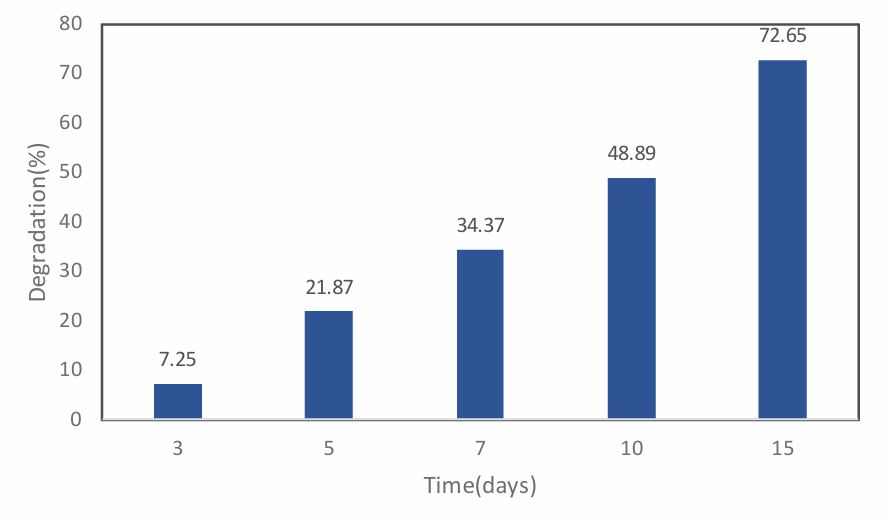
The fabricated hydrogel (HGEL-2) is mainly composed of CMC. The monomers of CMC are linked via glycosidic linkages which can be easily broken by various enzymes resulting in small polysaccharide chains. These chains are further broken down. Figure 5, it can be seen that, biodegradability of the hydrogel is increased with increasing time. From the above figure, it can also be seen that the minimum biodegradability of hydrogel is 7.25% at the time 3 days and the maximum biodegradability of hydrogel is 72.65% at 15 days. So, we can say that hydrogel is a biodegradable polymer.
Scanning electron microscopy (SEM) analysis of CMC and hydrogel
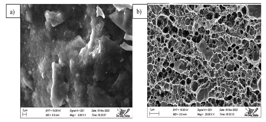
Scanning electron microscopy (SEM) offers several advantages for morphological analysis and has been widely used in fields such as metallurgy, natural and modified fibers, polymers, biomaterials, and composites [34]. Figures 6(a) and 6(b) show the surface structures of the synthesized CMC and the hydrogel derived from CMC, respectively, at 5000x magnification. As depicted in Figure 6, hydrogels with larger pore sizes exhibit denser network structures. It can be concluded that hydrogels with larger pores tend to have lower density due to the greater void volume fraction. Additionally, finer pores are visible in the hydrogel. The smooth, uniform appearance of the films indicates a compact structure with a clear porous surface. The analysis suggests that CMC, PVP, and their blends were chemically crosslinked with GA. The CMC surface appears rough, granular, and uniform, while the hydrogel SEM images reveal a greater number of pores and a more textured surface. These larger pores reduce diffusion resistance and enhance mass transfer, thanks to the increased internal surface area. This structure also provides a higher drug-delivery capacity, as confirmed by SEM analysis, which shows a significant surface area.
Antimicrobial study of hydrogel
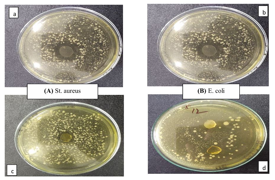
By measuring the inhibition zones that developed surrounding the hydrogel, the antibacterial activity was evaluated. Figure 7 displays the hydrogels' (HGEL-1, HGEL-2) antibacterial activity, and Table 2 contains the inhibition zone sizes (d in mm). Using the disc diffusion method and air as the negative control, the antibacterial effectiveness of hydrogels against Escherichia coli and Staphylococcus aureus was investigated.
| Bacterial cultures | Inhibition zone diameters (d in mm) for HGEL-1 | Inhibition zone diameters (d in mm) for HGEL-2 |
|---|---|---|
| St. aureus | 8 | 11 |
| E. coli | - | 11 |
The hydrogels possibly broke up the cell surface and made the outermost parts of the cells uneven. This stopped microbes from growing near the hydrogel samples (Figure 7). When bacterial cells came in contact with CMC, the -COOH group lost an H+ ion and was changed into a -COO− ion. The bacteria's pH was affected by the H+ ions, which split the cell wall. Also, the carboxylate ions disrupted up the work that was going on in the cells by connecting to things that were positively charged in the bacterial cells [35]. This behavior suggests that these hydrogels could enhance the antimicrobial effectiveness of antibiotics, making them suitable for use in wound healing applications.
Drug releasing study of hydrogel
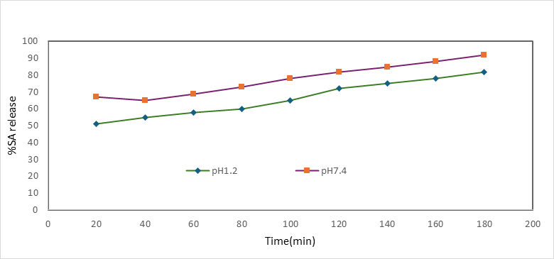
Water-soluble drugs that are trapped in hydrogels don't come out until water gets into the polymer networks, which makes the drug swell and dissolve. After this, the drug moves through the water channels and reaches the surface of the device [36]. In contrast to pH 1.2, where a total of 51% of the drug was released in 20 minutes, a total of 67% of the drug was released in 20 minutes. The SA release profile of the drug-loaded CMC/PVP hydrogel membrane (HGEL-2) in various release media, including pH 1.2, pH 7.4, and pure water, as shown in Figure 8. 67% of the drugs were released in 20 minutes, according to the hydrogel membrane's percent release profile at pH 7.4. At pH 1.2, only 51% of the drugs were released in the same amount of time. The release pattern shows that after three hours, 92% of the medicine was released at a pH of 7.4, and then 82% of the medicine was released at a pH of 1.2. Based on the results, we can guess that the pH 7.4 medium released more SA than the pH 1.2 medium and distilled water. You can connect this event to how the CMC/PVP hydrogel membranes swell at different pH levels (Fig. 8), where the swelling got bigger when the medium's pH went from mostly acidic to slightly basic. The carboxymethyl group in the CMC/PVP complex becomes ionized when the pH is high. Because of this, the –COO- groups push against each other, which makes the hydrogels that have already formed swell more and release more SA.
Conclusion
The crosslinked polymeric network structure of the hydrogel was successfully formed by the solution casting method. The properties like degree of swelling in different pH, water absorption, gel fractions, biodegradability, morphology and thermal behavior were examined for superabsorbent hydrogel. Those properties ensure the improved quality of hydrogels. The crosslinked three-dimensional network structure of hydrogels was confirmed by FTIR analysis. Scanning electron microscopy revealed the morphology of the hydrogels. TGA data gives an idea about the thermal stability of the superabsorbent hydrogel. The TGA curve shows the gradual decomposition of the hydrogel. It was also observed that crosslinking improved the thermal properties of hydrogels. In this work, glutaraldehyde plays an important role in producing a crosslinked polymeric network structure of superabsorbent hydrogel as a crosslinking agent. The use of non-toxic crosslinking agents is a current trend in the design of cellulose-based hydrogels, which improves the safety of the manufacturing process and the finished product. These hydrogels are especially appealing for targeted drug delivery because of their "smart" characteristics, which include reacting to physiological elements like pH. Furthermore, these hydrogels may soon be employed in wound healing applications due to their antimicrobial properties.
Ethical Approval
This work is an original contribution and has not been published previously in any form or language. The results are presented clearly, honestly, and without fabrication, falsification, or inappropriate data manipulation.
Consent to Participate
This study involved only laboratory-based experimental work. No human participants, animals, or living tissues were involved; therefore, consent to participate was not required.
Consent to Publish
The authors approve the publication of identifiable information in this journal or article, including photographs and/or text.
Declarations
- Author contributions
All authors contributed to the study conception and design. Material preparation, data collection and analysis were performed by Most. Arifa Sultana. All authors read and approved the final manuscript
- Funding
This research was funded by the University of Rajshahi through a university research project.
- Data availability
Not applicable
- Competing interests
Not applicable
- Acknowledgments
Most. Arifa Sultana gratefully acknowledges the financial support provided by the University of Rajshahi for conducting this research.
References
- 1.Thakur, S., Thakur, V. K., and Arotiba, O. A., (2018). History, classification, properties and application of hydrogels: An overview. Hydrogels: Recent Advances: 29-50.
- 2.Brannon-Peppas, L. and Peppas, N. A., (1991). Equilibrium swelling behavior of pH-sensitive hydrogels. Chemical Engineering Science, 46(3): 715-722.
- 3.Kloxin, A. M., Kloxin, C. J., Bowman, C. N., and Anseth, K. S., (2010). Mechanical properties of cellularly responsive hydrogels and their experimental determination. Advanced materials, 22(31): 3484-3494.
- 4.Peppas, N. A., Bures, P., Leobandung, W., and Ichikawa, H., (2000). Hydrogels in pharmaceutical formulations. European journal of pharmaceutics and biopharmaceutics, 50(1): 27-46.
- 5.Qiu, Y. and Park, K., (2001). Environment-sensitive hydrogels for drug delivery. Advanced drug delivery reviews, 53(3): 321-339.
- 6.He, B., Wan, E., and Chan-Park, M. B., (2006). Synthesis and degradation of biodegradable photo-cross-linked poly (α, β-malic acid)-based hydrogel. Chemistry of materials, 18(17): 3946-3955.
- 7.Treloar, L., (1952). The thermo dynamic study of rubber-like elasticity. Proceedings of the Royal Society of London. Series B-Biological Sciences, 139(897): 506-512.
- 8.Kumar, S. and Pandey, A. K., (2013). Chemistry and biological activities of flavonoids: an overview. The scientific world journal, 2013(1): 162750.
- 9.Kamath, K. R. and Park, K., (1993). Biodegradable hydrogels in drug delivery. Advanced drug delivery reviews, 11(1-2): 59-84.
- 10.Peppas, N. A., Huang, Y., Torres-Lugo, M., Ward, J., and Zhang, J., (2000). Physicochemical foundations and structural design of hydrogels in medicine and biology. Annual review of biomedical engineering, 2(1): 9-29.
- 11.Li, M., Cha, D. J., Lai, Y., Villaruz, A. E., Sturdevant, D. E., and Otto, M., (2007). The antimicrobial peptide‐sensing system aps of Staphylococcus aureus. Molecular microbiology, 66(5): 1136-1147.
- 12.Mitsumata, T., Suemitsu, Y., Fujii, K., Fujii, T., Taniguchi, T., and Koyama, K., (2003). pH-response of chitosan, κ-carrageenan, carboxymethyl cellulose sodium salt complex hydrogels. Polymer, 44(23): 7103-7111.
- 13.Hirsch, S. G. and Spontak, R. J., (2002). Temperature-dependent property development in hydrogels derived from hydroxypropylcellulose. Polymer, 43(1): 123-129.
- 14.Jaiswal, M. and Koul, V., (2013). Assessment of multicomponent hydrogel scaffolds of poly (acrylic acid-2-hydroxy ethyl methacrylate)/gelatin for tissue engineering applications. Journal of biomaterials applications, 27(7): 848-861.
- 15.Ricka, J. and Tanaka, T., (1984). Swelling of ionic gels: quantitative performance of the Donnan theory. Macromolecules, 17(12): 2916-2921.
- 16.Zhu, J. and Marchant, R. E., (2011). Design properties of hydrogel tissue-engineering scaffolds. Expert review of medical devices, 8(5): 607-626.
- 17.Kabiri, K., Omidian, H., Zohuriaan‐Mehr, M., and Doroudiani, S., (2011). Superabsorbent hydrogel composites and nanocomposites: a review. Polymer composites, 32(2): 277-289.
- 18.Taşdelen, B., Erdoğan, S., and Bekar, B., (2018). Radiation synthesis and characterization of chitosan/hyraluronic acid/hydroxyapatite hydrogels: Drug uptake and drug delivery systems. Materials today: proceedings, 5(8): 15990-15997.
- 19.Katono, H., Sanui, K., Ogata, N., Okano, T., and Sakurai, Y., (1991). Drug release off behavior and deswelling kinetics of thermo-responsive IPNs composed of poly (acrylamide-co-butyl methacrylate) and poly (acrylic acid). Polymer journal, 23(10): 1179-1189.
- 20.Shantha, K. and Harding, D., (2002). Synthesis and evaluation of sucrose‐containing polymeric hydrogels for oral drug delivery. Journal of applied polymer science, 84(14): 2597-2604.
- 21.Sri, B., Ashok, V., and Arkendu, C., (2012). As a review on hydrogels as drug delivery in the pharmaceutical field. Int J Pharm Chem Sci, 1(2): 642-61.
- 22.Ashley, G. W., Henise, J., Reid, R., and Santi, D. V., (2013). Hydrogel drug delivery system with predictable and tunable drug release and degradation rates. Proceedings of the national academy of sciences, 110(6): 2318-2323.
- 23.Park, C. H., Jeong, L., Cho, D., Kwon, O. H., and Park, W. H., (2013). Effect of methylcellulose on the formation and drug release behavior of silk fibroin hydrogel. Carbohydrate polymers, 98(1): 1179-1185.
- 24.Gerami, S. E., Pourmadadi, M., Fatoorehchi, H., Yazdian, F., Rashedi, H., and Nigjeh, M. N., (2021). Preparation of pH-sensitive chitosan/polyvinylpyrrolidone/α-Fe2O3 nanocomposite for drug delivery application: Emphasis on ameliorating restrictions. International Journal of Biological Macromolecules, 173: 409-420.
- 25.Palem, R. R., Rao, K. M., Shimoga, G., Saratale, R. G., Shinde, S. K., Ghodake, G. S., and Lee, S.-H., (2021). Physicochemical characterization, drug release, and biocompatibility evaluation of carboxymethyl cellulose-based hydrogels reinforced with sepiolite nanoclay. International Journal of Biological Macromolecules, 178: 464-476.
- 26.Sachan, N. and Bhattacharya, A., (2009). Modeling and characterization of drug release from glutinous rice starch based hydrogel beads for controlled drug delivery. International Journal of Health Research, 2(1).
- 27.Shaikh, H. M., Anis, A., Poulose, A. M., Al-Zahrani, S. M., Madhar, N. A., Alhamidi, A., and Alam, M. A., (2021). Isolation and characterization of alpha and nanocrystalline cellulose from date palm (Phoenix dactylifera L.) trunk mesh. Polymers, 13(11): 1893.
- 28.Jia, F., Liu, H.-j., and Zhang, G.-g., (2016). Preparation of carboxymethyl cellulose from corncob. Procedia Environmental Sciences, 31: 98-102.
- 29.Pal, K., Banthia, A. K., and Majumdar, D. K., (2007). Preparation and characterization of polyvinyl alcohol-gelatin hydrogel membranes for biomedical applications. Aaps Pharmscitech, 8(1): 21.
- 30.Nagasawa, N., Yagi, T., Kume, T., and Yoshii, F., (2004). Radiation crosslinking of carboxymethyl starch. Carbohydrate Polymers, 58(2): 109-113.
- 31.Mishra, R. K., Datt, M., and Banthia, A. K., (2008). Synthesis and characterization of pectin/PVP hydrogel membranes for drug delivery system. Aaps Pharmscitech, 9(2): 395-403.
- 32.Bonev, B., Hooper, J., and Parisot, J., (2008). Principles of assessing bacterial susceptibility to antibiotics using the agar diffusion method. Journal of antimicrobial chemotherapy, 61(6): 1295-1301.
- 33.Ali, A. E.-H., Shawky, H., Abd El Rehim, H., and Hegazy, E., (2003). Synthesis and characterization of PVP/AAc copolymer hydrogel and its applications in the removal of heavy metals from aqueous solution. European polymer journal, 39(12): 2337-2344.
- 34.Sikdar, B., Basak, R., and Mitra, B., (1995). Studies on graft copolymerization of acrylonitrile onto jute fiber with permanganate ion initiation system in presence of air. Journal of applied polymer science, 55(12): 1673-1682.
- 35.Kundukad, B., Schussman, M., Yang, K., Seviour, T., Yang, L., Rice, S. A., Kjelleberg, S., and Doyle, P. S., (2017). Mechanistic action of weak acid drugs on biofilms. Scientific reports, 7(1): 4783.
- 36.Singh, B., Chauhan, G., Sharma, D., and Chauhan, N., (2007). The release dynamics of salicylic acid and tetracycline hydrochloride from the psyllium and polyacrylamide based hydrogels (II). Carbohydrate polymers, 67(4): 559-565.
Cite this article
Journal
Sultana MA, Ahsan MS, Mondal A, Chowdhury A, Hassib MA, Alom A, Mondol AK, Molla MTH. Design and Performance of a Chemically Crosslinked CMC/PVP Hydrogel for Drug Release. JoSEP. 2026;3(1):145-152. doi: 10.62275/josep.26.1000023.
Rights and permissions
© 2026
Cited by (0)
Citing works are not available yet.
About this article
- Received
- 23 December 2025
- Revised
- 6 January 2026
- Accepted
- 7 January 2026
- Published
- 22 October 2026
- Journal
- Journal of Science and Engineering Papers
- Volume / Issue
- 3 / 1
- Pages
- 145-152
- DOI
- 10.62275/josep.26.1000023